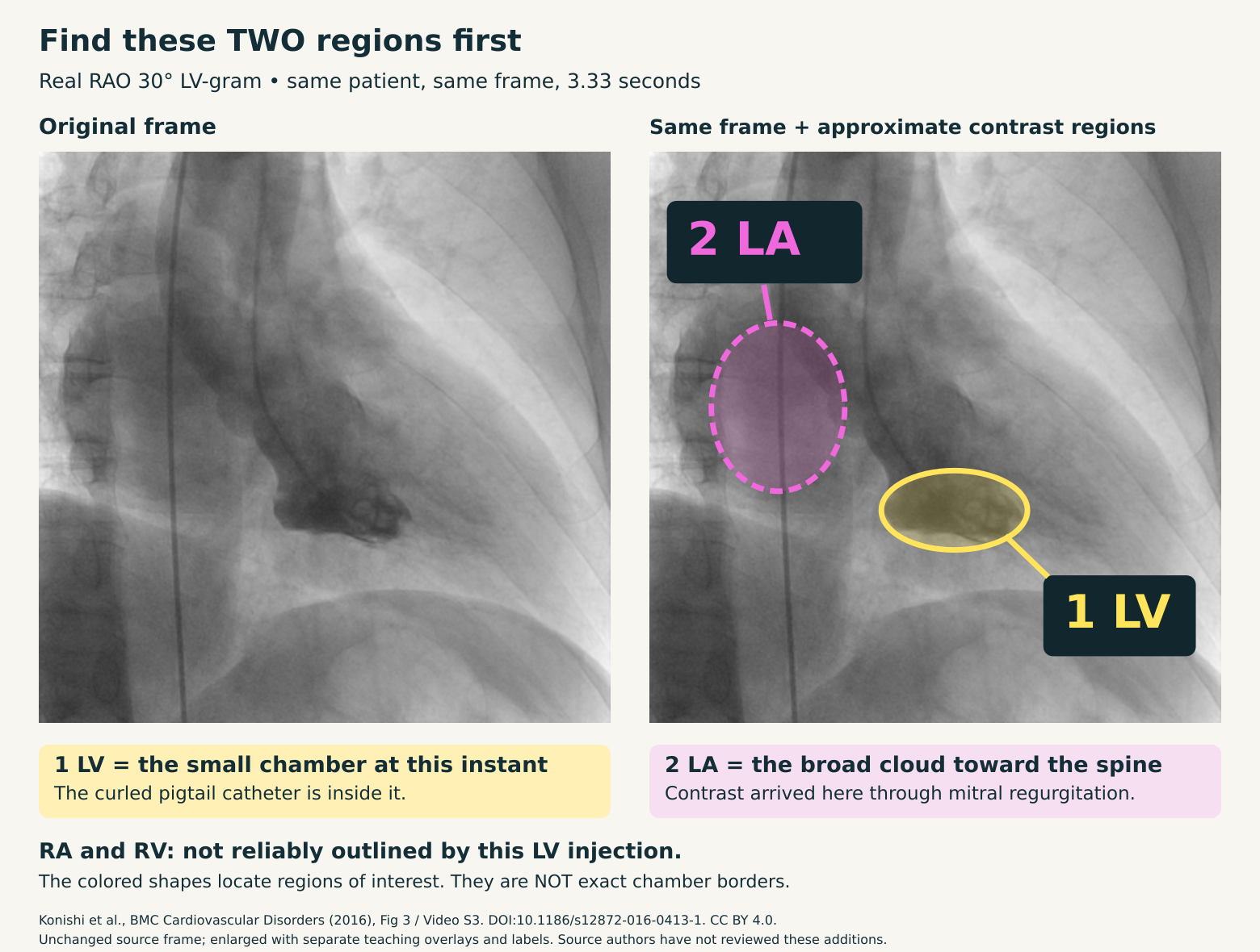
Find these two regions first.
Yellow = LV. Pink = contrast in the LA.
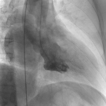
Exact same source frame in both panels. Colored shapes locate approximate contrast regions; they are not exact chamber borders.
RA and RV are not reliably outlined by this LV injection.
This is a projection with overlapping structures. You should not expect four separately visible, contrast-filled chambers in this shot.
Original source, attribution, and limits
Konishi et al., BMC Cardiovascular Disorders 16,234 (2016). Figure3 explicitly identifies RAO30; VideoS3 is the corresponding angiogram. Authors report grade3–4 MR. DOI:10.1186/s12872-016-0413-1.
© The Author(s),2016. CC BY4.0. Source video unchanged; a frame was extracted and enlarged with separate educational overlays and labels. Colored regions are approximate visual guides, not endocardial tracings or a quantitative measurement. The authors have not reviewed or endorsed the additions.
The systolic loop spans2.53–3.47seconds of the source; phase is identified by visible cavity reduction, with no synchronized ECG. This is a source-based educational example, not recalled examination content or a claim about which image an examination uses.